Why do they take a puncture from the knee joint? Don't be afraid of knee puncture - it doesn't hurt at all! Preparing the patient for the procedure
Puncture of the knee joint is a puncture into the cavity of the joint or joint capsules for the purpose of diagnosing any pathologies or treatment. This procedure is indispensable for many diseases of the knee joint, as it allows you to take joint fluid for subsequent laboratory tests.
Execution technique

The technique of performing the manipulation is not complicated at first glance and, taking into account the preparation of the patient, takes about 5-20 minutes. The puncture is carried out in the treatment room by the appropriate doctor, and in a sterile environment. The patient must first be notified of the need for this procedure and told how, what and why is being done, after which the patient gives his consent. Having laid the patient on the couch, the doctor asks to relax the leg, as tense muscles can interfere with the manipulation process.
Using a puncture, X-ray contrast agents can be injected into the joint to facilitate x-ray examination or medications.
The puncture is also used to extract the contents of knee joint cysts, which avoids surgical treatment.
It is more convenient to use one access to remove a sample of liquid for research, and another to administer medicinal substances. Immediately before the needle is inserted, the area is treated with iodine solution. Also, the procedure should be carried out under local anesthesia, this is either infiltration of the skin with a lidocaine solution, or the skin is irrigated with ethyl chlorine.
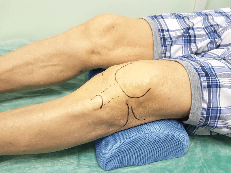
Important! The limb should be bent at the knee joint at an angle of approximately 10 - 20 degrees; to maintain the limb in this state and relax the muscles, a rolled up towel should be placed under the knee.
The technique and location of the injection depends on the purpose and purpose of the knee puncture. But there are only a few accesses, and mainly three are used:
- The injection using the first approach is made at a point 1 cm above the patella bone, the needle is inserted tangentially, almost parallel to the front surface of the thigh. Thus, a puncture of the lateral part of the superior inversion of the knee joint is performed.
- The second point is located below the patella. The doctor needs free hand displace the patella bone laterally - outwards, then the middle of the patella is located and along this line, half a centimeter below the lower edge of the patella, a puncture needle is inserted; it should be inserted slightly in the proximal direction. The correctness of execution should be judged by the feeling of the needle sinking, which occurs when it enters the joint cavity.
- The third way to puncture the knee joint involves inserting a needle at the lateral or medial edge of the patella slightly below the midline.
At the end of the manipulation, an aseptic bandage is applied to the injection site for several hours.
Indications
Knee puncture is prescribed for certain indications, and depending on the purpose, two types of knee puncture should be distinguished.
Diagnostic puncture of the knee joint cavity
During the procedure, part of the synovial fluid is removed from the joint cavity and sent for laboratory testing. This is of great diagnostic importance for many diseases associated with the knee joint, since the following factors can be detected in the joint fluid in various pathologies:
- blood;
- bacteria – causative agents of various infectious diseases;
- specific protein substrates characteristic of a certain pathological condition;
- atypical cells of bone or cartilage tissue characteristic of tumor processes.
Also, a diagnostic puncture is prescribed before arthroscopic operations or before the knee joint.
Therapeutic puncture of the knee
It is prescribed to remove pathological fluid from the joint cavity, for example, purulent exudate, blood or inflammatory exudative fluid. Also with therapeutic purpose. For example, antibiotics, anti-inflammatory substances, steroid drugs and other substances can be administered through a puncture. As a rule, for these purposes, the joint is punctured through several punctures, but this is individual and determined for a particular patient.

The therapeutic or diagnostic prescription is determined by the doctor, who is guided by the indications:
- acute inflammatory phenomena in the joint, with obvious accumulation of fluid;
- purulent arthritis;
- joint damage of an allergic nature;
- for diagnosis of chronic inflammation;
- for specific infectious diseases, for example, tuberculosis;
- injured joint with blood accumulation;
- planned before knee surgery.
As you can see, this procedure is prescribed for a wide range of indications, and if a doctor prescribes a puncture, then you should not refuse it.
Baker's cyst puncture
A Becker cyst is a formation located in the popliteal fossa, formed when effusion from the articular cavity of the knee joint into the subtendinous bursa associated with the joint and located in the popliteal fossa. This formation always occurs due to the effusion of fluid from the knee joint; the effusion can be of either inflammatory or non-inflammatory nature.
Education in itself does not cause obvious pain syndrome. The cyst is dangerous only when it reaches a significant size, as a result of which it can mechanically interfere with knee flexion or compress the underlying neurovascular bundle. When the nerves are compressed, tingling and numbness appear in the lower leg.
Also, such a cyst can act as a factor contributing to varicose veins veins of the lower leg, or their thrombosis. A cyst may rupture due to compression or when there is active and rapid accumulation of pathological contents in it. Rupture of a Baker's cyst threatens the penetration of contents into the intermuscular space of the leg, which will be accompanied by pain, swelling and hyperemia of the skin.

Baker's cyst puncture represents a conservative option for its treatment. Through puncture, pathological fluid is evacuated, both from the cyst itself and from the joint cavity, after which glucocorticosteroids are administered. But puncture, as a rule, gives a temporary effect, and relapses occur. In such cases, surgical treatment methods are resorted to. The operation is most suitable in in this case, but it is not recommended, for example, for children or due to contraindications, then they resort to puncturing the cyst.
Contraindications and complications
There are not many contraindications to this manipulation, but neglecting this can cause undesirable consequences and infectious complications. Knee puncture is contraindicated in the following cases:
- For skin lesions in the joint area, for example, with psoriasis.
- It is also not advisable to perform manipulation on traumatically damaged skin.
- But infectious lesions are a categorical contraindication.
- Impaired blood clotting is also considered a serious contraindication.
If there are such disorders, the patient must be prepared for the procedure by administering special medications. At first glance, the procedure seems quite simple from the point of view of the technique, but it should only be carried out by a specialist doctor, since careless insertion of the needle or failure to comply with sterile conditions can lead to undesirable consequences and complications.
Often these are hematomas at the injection site due to damage to blood vessels; damage to the articular cartilage and synovial membrane is possible, which will take a long time to heal. A serious complication is purulent arthritis, which is inflammation of the joint due to infection.
Rashes and allergic reactions of the patient to the administered drugs are also possible. If swelling or swelling of the joint appears or if the temperature rises after the procedure, you should immediately consult a doctor.
Important! To perform the operation, you must follow all the rules of sterility, use disposable syringes, only sterile gloves, swabs and needles.
It is also necessary to sanitize your hands and the site of the procedure before the procedure. With a competent and responsible attitude to the procedure on the part of both the doctor and the patient, complications can be avoided, and the procedure is successful, bringing only a positive effect.
Prices and reviews about the procedure
The price for a puncture of the knee joint depends on the purpose of its implementation and on the institution where the procedure takes place. According to the compulsory medical insurance policy, if a procedure is necessary for treatment or diagnosis, it is carried out free of charge. On average, in Russia the price of the procedure ranges from 1 – 2 thousand rubles. Reviews from people who have undergone a puncture are full of positive assessments.
I have been running for a long time; I recently had a knee injury, and the pain did not go away for a long time. I visited the doctor and prescribed a puncture to take tests. I was a little afraid, I thought it would hurt, but under anesthesia you don’t feel anything, don’t be afraid. Fortunately, the doctor turned out to be competent, and everything went without complications.
Vasily, 21 years old.
The child began to complain of pain under his knee, and a lump came out. The hospital said that it was a Baker's cyst and that a puncture was needed, since the child could not undergo surgery. They did it successfully, the pain went away, everything is fine, the child is running. They said relapses are possible, but we hope it will work out.
Elena, 43 years old.
I had to experience it myself. My knee hurt, they said there was fluid in the joint, I needed to do a puncture, then they would be more specific. I was afraid it would hurt, I watched a video of how they do it and calmed down. And in fact, everything is not so scary, they did it afterwards and it didn’t hurt much.
Indications. Joint puncture is used for diagnostic and therapeutic purposes to determine the nature of the contents in it (effusion, blood), remove this content from the joint cavity and administer antiseptic solutions or
antibiotics. For puncture, a 10-20 gram syringe equipped with a thick needle is used; less often, a thin trocar is used (for the knee joint). Before puncturing the joint, prepare the instruments, the surgeon’s hands and the surgical field, as for any surgical intervention.
Anesthesia - local novocaine anesthesia. To puncture a joint, it is recommended that before inserting a needle, move the skin in this place to the side with your finger. This achieves curvature of the wound channel (where the needle passed) after the needle is removed and the skin falls into place. This curvature of the wound channel prevents the contents of the joint from leaking out after the needle is removed. The needle is advanced slowly until a sensation appears indicating a puncture of the joint capsule. After the operation is completed, the needle is quickly removed and the puncture site is sealed with collodion or adhesive tape. The limb must be immobilized with a plaster cast or splint.
10.11.1. Puncture of the joints of the upper limbs
Shoulder puncture
If there are appropriate indications, puncture of the shoulder joint can be performed both from the anterior and posterior surfaces. In order to puncture the joint from the front, the coracoid process of the scapula is probed and a puncture is made directly under it. The needle is advanced posteriorly, between the coracoid process and the head of the humerus, to a depth of 3-4 cm. The puncture of the shoulder joint from behind is carried out through a point located below the posterior edge of the apex of the acromial process, in the fossa formed by the posterior edge of the deltoid muscle and the lower edge m. supraspinatus. The needle is passed anteriorly towards the coracoid process to a depth of 4-5 cm (Fig. 10-8 a).
Rice. 10-8. Puncture of the shoulder (a), elbow (b) and wrist (c) joints
Elbow puncture
The arm is bent at the elbow joint at a right angle. The needle is inserted from behind between the lateral edge olecranon and bottom edge epicondilis lateralis humeri, directly above the head of the radius. The superior inversion of the joint is punctured above the apex of the olecranon process, moving the needle down and anteriorly. Puncture of the joint along the medial edge of the olecranon is not used due to the risk of damage to the ulnar nerve (see Fig. 10-8 b).
Wrist puncture
Because joint capsule Since the palmar surface is separated from the skin by two layers of flexor tendons, a more accessible place for puncture is the dorsal radial surface. The injection is made on the dorsal surface of the joint area at the intersection of the line connecting the styloid processes of the radius and ulna with the line that is a continuation of the second metacarpal bone, which corresponds to the space between the tendons m. extensor policis longus et m. extensor indicis(see Fig. 10-8 c).
10.11.2. Puncture of the joints of the lower extremities
Knee joint puncture
Indications: hemarthrosis, intra-articular fractures.
Technique. Treat the skin with alcohol and iodine. On the outside of the patella, the skin is anesthetized with a 0.5% novocaine solution. The needle is directed parallel to the posterior surface of the patella and penetrates the joint. A syringe is used to evacuate blood from the joint. If there are intra-articular fractures, after removing the blood, 20 ml of a 1% novocaine solution is injected into the joint to anesthetize the fracture site (Fig. 10-9).
 Rice. 10-9. Knee joint puncture
The puncture of the superior inversion of the knee joint is most often performed at the lateral edge of the base of the patella. The needle is advanced perpendicular to the axis of the thigh under the tendon of the quadriceps muscle to a depth of 3-5 cm. From this point the knee joint can be punctured. In this case, the needle is directed downward and inward between the posterior surface of the patella and the anterior surface of the epiphysis of the femur.
No complications are observed if technique and asepsis are observed.
Puncture hip joint
Puncture of the hip joint can be performed from the anterior and lateral surfaces. To determine the injection point, use the established joint projection diagram. To do this, draw a straight line from the greater trochanter to the middle of the Pupart ligament. The middle of this line corresponds to the femoral head. At the point established in this way, a needle is inserted, which is carried perpendicular to the plane of the thigh to a depth of 4-5 cm until it reaches the femoral neck. Then the needle is turned slightly inward and, moving it deeper, it penetrates into the joint cavity (Fig. 10-10). Puncture of the upper part of the joint can also be performed above the apex of the greater trochanter, passing the needle perpendicular to the long axis of the femur. As the needle penetrates the tissue, it rests on the femoral neck. By giving the needle a slightly cranial direction (up), they enter the joint.
 Rice. 10-10. Puncture of the hip joint.
a - diagram of puncture of the hip joint; b - technique of puncture of the hip joint
Ankle puncture
Puncture of the ankle joint can be performed from the outer or inner surface. To determine the puncture point, use the joint projection diagram (Fig. 10-11 a, b). The puncture point on the outer surface of the joint is 2.5 cm above the apex of the lateral malleolus and 1 cm medially from it (between the lateral malleolus and m. extensor digitorum longus). The puncture point along the inner surface of the joint is located 1.5 cm above the medial malleolus and 1 cm medially from it (between the inner malleolus and m. extensor halucis longus). After soft tissue anesthesia, a joint puncture is performed at the targeted point by inserting a needle between the talus and ankle. Fluid or blood is removed from the joint cavity, and if necessary, a medicinal substance (antibiotics, antiseptics) is administered.
 Rice. 10-11. Puncture of the ankle joint.
a - diagram of the projection of the ankle joint; b - technique of puncture of the ankle joint
When treating diseases of the musculoskeletal system, modern medicine uses an integrated approach. Combinations medicines different groups are well combined with surgical interventions, mechanotherapy and physiotherapy, folk remedies. Prosthetics, arthroscopy, and puncture are widely used today to restore movement functions.
What is a joint puncture
Puncture of any joint is the name of the procedure according to which the intraarticular membrane is pierced through the skin with a needle and penetrates into the cavity. The puncture is performed for therapeutic or diagnostic purposes.
The puncture technique (also called arthropuncture or puncture) requires professionalism and accuracy from the specialist so as not to damage the nerve endings. IN otherwise this can cause severe pain and, much worse, significant restriction in movement.
For this reason, such a procedure is carried out exclusively by a specialist (surgeon and/or traumatologist) in a hospital setting.
A significant role in this treatment is played by the individual anatomical structure of the joints, as well as the location of the surrounding tissues of the individual patient. For example, on the forearm, large vessels and nerves are located rather superficially; for this reason, arterial puncture of the elbow joint is performed with special care. Well-developed muscles of the limbs, on the one hand, protect the joints from injury, on the other, prevent rapid access to them. For example, it involves a difficult technique and involves the use of special devices.
Indications for referral for the procedure
This procedure is quite common in surgery and traumatology. For diagnostic and therapeutic purposes, puncture of the knee joint is prescribed to the patient in the following cases:
Important for the patient before administration of various medications, inform in advance about allergies or other medications taken in order to avoid drug side effects.
Causes and symptoms of synovitis disease
 If pain is localized in the knee joint, you should immediately consult a doctor. The fact is that these may be the initial signs of synovitis. This disease can occur in at different ages. It occurs due to allergic reactions, arthritis, and injuries. Near the knee joint Fluid begins to accumulate in significant quantities, due to which any movement of the leg causes acute pain. There are main signs by which this disease can be identified:
- The knee grows in size.
- There is a bluish or red appearance around the joint.
- Acute pain occurs.
- In rare cases, increased body temperature.
A puncture will help cope with the disease. The doctor removes the unnecessary fluid, and then an antibiotic is injected into the leg. The patient immediately feels relief. The disease is dangerous because if the problem is not eliminated in a timely manner, the knee joint can completely collapse, thereby affecting the meniscus. To cope with this trouble, long-term treatment and surgical intervention will be required.
Stages of performing joint puncture
The procedure technique is identical for each joint. Let's talk about performing this procedure on the knee joints.
Due to the simple anatomical structure, knee puncture is simpler. The technique of puncturing the knee joint consists of several stages:

Possible consequences of treatment
 If all the rules are followed, as a result of the puncture, positive dynamics in the treatment process are observed. When removed from a joint Excessive exudate eliminates painful sensations, since intra-articular pressure decreases. Upon implementation medicines The body's inflammatory reactions are removed from the knee, pain and swelling go away, and regeneration processes increase. Just a day later, the patient can begin rehabilitation treatment, including physiotherapeutic procedures and exercise therapy.
The use of inappropriate instruments (diameter and size of the puncture needle is too large) can result in damage to ligaments, bone structures or muscle nerves (puncture of the elbow joint).
From time to time, local irritation of the skin surface in the area of iodine treatment may occur. This is actually why it is recommended to use modern antiseptics. Regular administration of hormonal medications may lead to destruction of intra-articular cartilage.
It is extremely important to carefully follow the instructions for use of each pharmaceutical drug. For example, a combination of certain medications with concomitant hypertension, diabetes mellitus, kidney or liver damage can significantly aggravate the patient’s condition.
A puncture of the knee joint by a professional will not only help you resume a normal lifestyle, but also speed up your recovery.
Negative consequences may be accompanied by:
- joint infections;
- rupture of the synovial membrane.
If the knee is swollen, the temperature has risen, or throbbing or “burning” pain is felt, you should immediately consult a doctor, since such signs indicate infection has penetrated into the joint. Similar symptoms may not appear immediately, and a few days after completion of the procedure. You can avoid infection by following the puncture technique and all precautions. Many medications that are intended to be administered into the knee joint are deliberately prepared in disposable syringes.
If the membrane ruptures, acute pain is felt instantly as fluid begins to flow into the soft tissues of the body. Fortunately, such a complication is very rare. Allergic reactions are also possible, not to the puncture itself, but to substances that are part of the medications prescribed for therapy. That is why the doctors are not to blame in this case.
Contraindications
Not so often, but there are still situations when it would be better to refuse a puncture. This decision is made when the procedures themselves may harm the patient more than they can benefit. Such cases include the following indicators:
- Increased blood clotting.
- Ulcers or wounds are observed on the surface of the skin.
- The patient suffers from psoriasis.
- The patient is diagnosed with skin cancer.
 In other cases, this treatment can be safely prescribed and carried out. The most important thing is to find a competent specialist who will be able to carry out all the manipulations correctly.
On various forums you can often see the question: “If the joint in the knee hurts, is it necessary to perform a puncture or can it be avoided?”. Doctors are confident that without this procedure it is very difficult to cure synovitis. To begin with, it is necessary to remove excess fluid, and then prescribe the use of ointments and antibiotics.
Actively influence the course of the pathological process musculoskeletal system puncture of the knee joint helps. According to indications, puncture of the hip joint, as well as other affected joints, is also performed.
What is a knee puncture?
The treatment is performed by inserting a special needle into the cavity through special points. To do this, it is necessary to pierce the skin, muscle layer, tendons and joint membrane. This procedure can only be performed by a qualified traumatologist in a specially equipped office or hospital.
Types of punctures
Depending on the purpose, therapeutic and diagnostic manipulations are distinguished.
Therapeutic puncturing
Injecting the drug into the joint cavity is the most effective way effects on inflammatory and degenerative processes of the musculoskeletal system. The advantage of the method is to minimize side effect drugs, as well as their direct effect on tissue. To achieve the desired effect, puncture of the ankle joint and other damaged joints is performed using the same technique.
For therapeutic purposes, the following is injected into the joint cavity:
- antibiotics;
- anti-inflammatory steroidal and non-steroidal drugs;
- chondroprotectors;
- saline solution for rinsing;
- hemostatics;
- oxygen.
Treatment with direct injection of drugs into the joint area allows the most effective influence on the pathological process.
The joint is punctured in order to extract from it:
- excess synovial fluid;
- blood;
- purulent contents.
Removal of pathological exudate by puncture promotes rapid recovery of the patient.
Diagnostic puncturing

A puncture will help clarify the nature of the inflammatory process, as well as recognize traumatic injuries to the joint. Taking fluid allows you to determine the pathogen that caused the infectious process. The arrival of contrast agents makes it possible to diagnose traumatic lesions of the meniscus and articular surfaces.
punctuation points
Correctly selected points for puncture of the knee joint will ensure painless penetration into the cavity without damage important entities. The reference point for this is the patella. Places should be punctured from the outer or inner side under the lower edge or below the midline by approximately 5 mm. It is permissible to perform a puncture 1 cm above the upper edge of the patella. The needle is located parallel to its inner surface.
Indications for use
Puncture of the affected knee is necessary in the following situations:
- accumulation of blood in the joint cavity (only its mechanical removal can restore functioning);
- damage to the synovial membrane as a result of traumatic impact and excessive accumulation of intra-articular fluid;
- in the presence of infection of the joint capsule with the formation of purulent contents;
- pain relief through the administration of local anesthetics, manipulation of the reduction of a dislocated knee joint;
- deforming processes with degeneration of the cartilage surface and the presence of pain in the affected area;
- Baker's cyst (its contents are removed);
- progressive adhesive process.
Complex traumatic injuries require the introduction of contrast agents through puncture for accurate diagnosis of the pathological process.

Puncture of the elbow joint and other joints has similar indications.
Contraindications for use
The procedure cannot be performed if the patient has:
- violation of the integrity of the skin in the projection of the planned manipulation;
- psoriatic formations and rashes of other origin in the knee joint;
- general intoxication with a feverish state.
You will need to refrain from performing a puncture in cases of diseases accompanied by reduced activity of the blood coagulation system (hemophilia, hemorrhagic vasculitis).
How is the puncture performed?
The technique for performing joint (knee) puncture includes the following steps:
- Giving the patient a comfortable position (lying on his back, a soft cushion is located under the knee or ankle to relax the leg muscles).
- Disinfection skin knee joint with a 5% alcohol solution of iodine, and then with a 70% ethyl alcohol solution.
- Selection of the necessary needles: their diameter for administering medications and oxygen should be 1 mm, and for pumping out liquid – no more than 2 mm.
- Depending on the purpose of the puncture, prepare a syringe with a volume of 5 to 20 ml.
To prevent pain for the patient during the procedure, local anesthesia is performed, for which the skin in the puncture area is injected with Novocaine or Lidocaine.

The skin fold at the puncture site shifts to bend the channel of the puncture needle. This prevents the possible entry of an infectious pathogen into the joint cavity.
After the procedure is completed, the injection site is covered with a pressure bandage.
The shoulder joint and other affected joints are punctured in the same sequence.
Standard puncturing technique
Puncturing by traditional technology involves inserting a needle into a slit hole determined by palpation. It is located between the greater trochanter of the femur and the area of the posterior edge of the patella. When entering the cavity, a feeling of falling into the void appears, this signals that the manipulation has been carried out correctly.
When the needle hits the bone surface, you will need to make movements until you feel a sinking feeling.
Upper inversion puncture
The formation of a large amount of synovial fluid makes it possible to puncture the joint using the superior inversion technology. Use your palm to press on the lower part of the patella and move it upward. Pathological effusion also moves upward. The needle is passed into the joint cavity through the quadriceps femoris.
Puncture of the inferior inversions
The puncturing technology using this method involves inserting a needle into the area of the lower part of the patella in the direction of the articular cavity. The knee is shifted by pressing the hand from top to bottom.
Complications and side effects

Adverse consequences of puncture of the knee joint occur only when the technology is violated: a correctly performed manipulation is not accompanied by severe painful sensations. Ignoring the rules of asepsis and antisepsis can cause infection of joint tissues.
When an intolerance reaction to an anesthetic or therapeutic agent develops, a rash appears in the form of urticaria. The most common causes of allergic reactions are the following drugs:
- Novocaine or Lidocaine;
- antibiotics;
- non-steroidal anti-inflammatory drugs.
The puncture procedure of the joint is performed with caution if the patient is diagnosed with diabetes mellitus, due to the possibility of the formation of a non-healing trophic ulcer at the puncture site.
For patients suffering from bronchial asthma, intra-articular administration of non-steroidal anti-inflammatory drugs is contraindicated due to the high likelihood of developing bronchospasm and anaphylactic shock.
There is a possibility of mechanical damage to the synovial membrane up to its complete rupture. Complications will not arise if the joint puncture technique is strictly followed.
Before the procedure, you should collect an allergy history and find out which drugs the patient cannot tolerate. This will prevent complications and select the most effective medications.
Puncture of the knee joint is one of the most effective therapeutic and diagnostic procedures for pathological conditions and injury. Compliance with puncture technology and its timely implementation provides a quick positive effect. The intervention should only be performed by a qualified specialist in the surgical department.
All materials on the site were prepared by specialists in the field of surgery, anatomy and specialized disciplines.
All recommendations are indicative in nature and are not applicable without consulting a doctor.
The knee joint is very susceptible to a variety of diseases, both traumatic and inflammatory. Constant load and its use in Everyday life make him vulnerable, and surgical treatment is often the only way to restore his mobility and functionality.
Patients who need knee surgery include young people who have suffered injuries, professional athletes, older people with degenerative processes, and even children and adolescents who need to have a sprain corrected.
 One of the most informative diagnostic procedures performed for pathology of the knee joint is its puncture. It can also be a therapeutic manipulation. Puncture of the knee joint should only be performed by a qualified specialist, because the joint has an extremely complex structure, and if the procedure is not followed, serious complications can occur.
A puncture is a puncture of a joint with a long needle, through which either medicine is injected or pathological contents are removed. Puncture of the joint capsule requires pain relief, but it can be done on an outpatient basis.
The purpose of the puncture is to diagnose pathological changes or treat a diagnosis established through non-invasive methods. After a therapeutic puncture, the patient feels significant relief, and if diagnosed, the doctor receives a sufficient amount of information to develop further tactics to combat the pathology.
Indications and contraindications for surgery
 Indications for puncture of the knee joint are:
- Intra-articular hematoma;
- Arthrosis, arthritis and other rheumatological diseases;
- Inflammation of the synovial lining of the joint after injury;
- Collection of material for cytological or histological analysis;
- Accumulation of pus in the joint cavity;
- The need for pain relief when eliminating a dislocation;
- Administration of drugs into the joint;
- Suspicion of tumor growth;
- Infectious arthritis (including tuberculosis).
Removing pathological contents from the joint reduces the pressure inside it, so that the patient quickly feels significant relief. In addition, the manipulation prevents further damage to the articular structures, their inflammation and necrosis, which threaten complete immobilization.
Injecting medications directly into the joint is an excellent alternative drug treatment, since the drug acts directly in the damaged area, there is no need to use high doses of medications that negatively affect the liver and kidneys, and side effects at the same time minimal.
Reduction of a dislocation is a very painful procedure that requires adequate pain relief. In this case, the trauma surgeon can inject the anesthetic directly into the knee joint to achieve the best effect.
In some cases, it is not possible to establish an accurate diagnosis using ultrasound and other non-invasive procedures. Puncture in diagnostically difficult cases makes it possible to obtain joint tissue and its contents, as well as conduct histological and cytological examinations, and determine the sensitivity of the exudate to antibiotics. In such cases, a diagnostic puncture is prescribed, which, at the same time, can become therapeutic.
Diagnostic puncture may be accompanied by the introduction of air or contrast into the joint, which facilitate the search and determination of the nature of the pathological focus. The contrast agent is clearly visible during X-rays and CT scans, which complement joint puncture.
Knee puncture may be contraindicated for several reasons:
- Bleeding disorders;
- Allergy to used anesthetics;
- Wound infection, pustular skin lesions, dermatitis in the area where the puncture is supposed to be performed;
- Psoriasis in the joint area.
In case of inflammatory changes in the skin, suppuration and infection, puncture carries a high risk of infection entering the joint cavity with the development of acute arthritis, so the procedure is postponed until the skin lesions are completely eliminated.
Preparation for puncture of the knee joint
Puncture of the knee joint is an outpatient procedure that does not require special and lengthy preparation, and in some cases the operation is indicated urgently, when there is simply no time for tests. At the same time, the patient must undergo a minimum of examinations, including general clinical blood and urine tests, determination of blood clotting, X-rays, CT or ultrasound of the joint.

Before the puncture, the doctor must find out whether there is an allergy to anesthetics, check whether the patient has undergone any interventions in the past and how they proceeded, and whether medications are constantly taken.
In the case of a planned puncture, anticoagulants and blood thinners are canceled, and if the operation is performed urgently, then measures are taken to prevent bleeding, since it will not be possible to cancel medications already taken.
For a planned puncture, the patient comes to the surgeon at the appointed time. Clothes and shoes should be comfortable, and it is better to take care of the journey home in advance, as pain is possible after the procedure. It is better not to drive behind the wheel immediately after a puncture of the joint cavity.
In case of inflammation of the joint elements of an infectious nature, antibiotics may be prescribed before puncture. wide range actions. Rheumatology patients take glucocorticosteroids to reduce swelling of the soft tissues of the joint.
A trauma surgeon from either a regular public hospital or a private medical center can perform a puncture, but the presence of appropriate qualifications and experience is considered a prerequisite. Cost in this case should not be the determining factor, because a highly qualified specialist can perform a free operation.
Technique for puncture of the knee joint
For proper puncture of the articular cavity, knowledge of the anatomy of the knee joint and the options for the passage of blood vessels and nerves in it is very important. Technical inaccuracies can cause serious complications, so the manipulation should only be performed by a highly qualified surgeon.
In the knee there is contact between the articular surfaces of the two large bones legs - tibial and femoral. To cushion and provide multidirectional movements, it is equipped with cartilage layers (menisci), as well as many ligaments that provide the highest endurance of the joint to significant mechanical loads.
The patella is located in front of the joint, protecting the internal contents from direct injury. It is quite mobile and can be easily moved by the surgeon’s hand in different directions, making it possible to obtain the shortest and safest paths for the puncture needle.

puncture of the knee joint
The technique for performing a puncture of the knee joint is quite simple:
- Selecting an access point;
- Penetration into the joint;
- Removing contents, taking tissues for analysis, etc., depending on the purpose of the procedure;
- Removing the needle and applying a bandage.
Pursuing a specific therapeutic or diagnostic goal, the surgeon chooses optimal points for puncture of the knee joint, which can be:
- In the upper outer or inner corners of the patella;
- The lower corners on the outer or inner side of the patella.

puncture points of the knee joint
The most commonly used access is in the upper outer corner, when the needle is injected one and a half to two centimeters outward and downward from the top of the patella. There is no cartilage in this place and the joint is not covered by muscles, so only skin, subcutaneous fat and the joint capsule itself are in the path of the needle.
After puncturing the skin, the needle moves at a right angle to it in a horizontal plane and passes behind the patella. The puncture depth is no more than 2.5 cm. This method of puncture is considered not only the simplest, but also the safest for the patient.
If it is impossible to puncture in the upper outer part of the patella, other ways are chosen:
- Nizhnenaruzhny- one and a half to two centimeters retreat from the edge of the patella outward and downward, placing the needle behind the patella and penetrating into the tissue to a depth of 2.5 centimeters;
- Inferomedial approach- one and a half to two centimeters down and to the side from the patella;
- Superomedial- moving 1.5-2 cm outward and upward, the needle moves to the central part of the patella to a depth of no more than 2.5 cm.
Having chosen the optimal point for puncturing the knee joint, the doctor treats the puncture site with antiseptic solutions; if there is hair, it must be removed in advance. Before puncture, local anesthetics (usually novocaine) are injected into the soft tissues, the patient is placed on his back with his legs straightened, and the punctured knee should be as relaxed as possible.
For puncture, needles 4-5 cm long are used. The moment when the needle has penetrated the joint, there is a feeling of failure, at which the surgeon takes special care, stopping further active advancement of the needle deeper.
If the purpose of the puncture is to remove pathological contents, then after penetrating into the joint cavity, the surgeon sucks out what is there (blood, pus) with a syringe, evaluates the resulting exudate visually and can send it for laboratory testing.
After evacuating the contents of the joint, its cavity is washed with a sterile solution of sodium chloride. It is possible to administer antibiotics and hormones immediately after emptying the joint. Lidocaine or novocaine is also injected into the joint after the puncture for pain relief.
When all the actions planned during the puncture are completed, the doctor removes the needle, treats the puncture site with an antiseptic, applies a sterile napkin and a tight bandage to prevent the formation of a hematoma.
After puncture of the knee joint, the patient can go home; there is usually no need for hospitalization unless there are severe injuries, in which puncture is one of the stages of hospital treatment.
After a puncture of the knee joint, there are usually no complications, the procedure is well established and does not pose any difficulty for a trauma surgeon, however, in rare cases, patients may encounter some unpleasant consequences, including infection with the development of purulent arthritis, intra-articular hematoma due to bleeding disorders or taking anticoagulants.
Complications may be associated with a violation of the puncture technique, insufficient experience of the surgeon, concomitant unfavorable background in the patient himself (diabetes, obesity, elderly age). In rare cases, an allergy to anesthetic drugs may occur.
Video: technique for puncture of the knee joint