Longitudinal section of the root, pencil drawing. Practical work “Structure of a cross section of a stem. Internal structure of a plant root
Tasks
9th grade
Dear Guys!
When answering questions and completing tasks, do not rush, as the answers are not always obvious and require the use of not only biological knowledge, but also general erudition, logic and a creative approach. Good luck in your work!
Part 1. You are offered test tasks that require a choice only one
answer out of four possible. The maximum number of points that can be scored is 50 (1 point for each test task). The answer index you think
the most complete and correct, indicate in the answer matrix.
The causative agent of cholera in cell shape is:
a) bacillus;
b) vibrio;
c) spirilla;
d) coccus.
Ferns have:
a) leaves, stems and roots, but do not have flowers or seeds;
b) leaves and roots, but do not have stems, flowers or seeds;
c) leaves, stems, roots and seeds, but do not have flowers;
d) stems and roots, but do not have leaves, flowers or seeds.
From a fern spore a shoot grows,
which contains:
a) antheridia;
b) archegonia;
c) both antheridia and archegonia;
d) does not contain antheridia and archegonia, since
is a sporophyte.
The figure shows a longitudinal section of a plant root. The number 5 on it indicates:
a) root hairs;
b) lateral roots;
c) adventitious roots;
d) hyphae of mycorrhizal fungi.
Adapting to life on land, higher plants did not immediately lose the mobility of male gametes. Of the listed plants, gametes lack flagella in:
a) pine trees;
b) cycad;
c) cuckoo Lena;
d) moss.
Of the listed functions of integumentary tissue, the most important for the first land plants was:
a) photosynthetic;
b) mechanical;
c) conductive;
d) protection against water loss.
AND  The thorn shown in the figure is:
a) modification of the leaf;
b) modification of the stipule;
c) modification of the shoot;
d) growth of integumentary tissue.
Wind-pollinated trees typically bloom in the spring before their leaves emerge in order to:
a) do not compete with pollinating insects;
b) more pollen fell on the stigmas;
c) photosynthesis did not interfere with pollination;
d) their flowers were clearly visible.
Water, with minerals dissolved in it, carries out an ascending path in the leaf in the following sequence:
a) stomata – leaf pulp cells – vessels;
b) vessels – cells of the leaf pulp – stomata;
c) sieve tubes - vessels - cells of the leaf pulp;
d) sieve tubes – leaf pulp – stomata.
Having studied the anatomical structure of the leaf of a flowering plant, the biologist discovered that its structure lacks stomata. This observation allowed him to conclude that this leaf belongs to a plant that grew:
a) in a body of water;
b) in a moderately humid forest;
c) in the meadow;
d) in a dry sandy place.
A plant whose underground partare not
onion:
a) lily;
b) garlic;
c) gladiolus;
d) narcissist.
The structure of the stem of monocotyledonous plants lacks:
a) bast;
b) peel;
c) cambium;
d) wood.
Coffee is a plant of the madder family. Evergreen or deciduous trees and shrubs. The flowers are 5-7-membered, with a funnel-shaped white corolla, fragrant. Fetus:
a) berry;
b) drupe;
c) cynarrhodium;
d) multidrupe.
The sundew can exist normally for a long time, without “feeding” on insects, under the following conditions:
a) in high light conditions;
b) in the presence of available forms of nitrogen in the habitat;
c) in the presence of available forms of sodium in the environment;
d) at low soil acidity values.
Schoolchildren sowed beets in the school plot at the end of April. The seeds have sprouted. And at the end of May, frosts were recorded in this area, when night temperatures dropped to -7ºC for several days. It can be assumed that this will lead to:
a) the formation of juicier and larger fruits;
b) the formation of only vegetative organs, because beets are a biennial plant and produce flowers and fruits in the second year;
c) the appearance of juicier and larger root crops, because low temperatures stimulate the rapid accumulation of sugars in underground organs;
d) flowering of beets in the first year.
In the 19th century in Germany, when gas mains supplying street lights broke, trees growing near the accident site shed their leaves even in summer. This effect is explained by the presence in the composition of the illuminating gas:
a) ethanol;
b) ethane;
c) ethylene;
d) acetylene.
Among bivalves there are predatory representatives. In predatory bivalves, compared to filter feeders, the following structural changes are observed:
a) the shell disappeared;
b) there are no closing muscles;
c) no siphons;
d) gills are reduced.
Toothless:
a) there is only a radula;
b) there is both a radula and a lock;
c) there is only a lock;
d) there is neither a radula nor a lock.
Coprophages are:
a) dung beetles;
b) gravedigger beetles;
c) leaf-cutter ants;
d) dead-eating beetles.
On the mesothorax of the housefly are:
a) three pairs of legs and one pair of wings;
b) one pair of legs and one pair of wings;
c) one pair of legs and two pairs of wings;
d) one pair of legs.
When insects emerge from the pupa, their wings spread out. due to:
a) pumping air into the wing;
b) gravity;
c) pumping hemolymph into the wing;
d) muscle contractions.
Among the food items used by aquarists, oligochaete worms include:
a) bloodworm;
b) tubifex;
c) mealworm;
d) Artemia.
In the figure, numbers 1–3 indicate coral structures: 
a) 1 – fringing reef, 2 – barrier reef, 3 – atoll;
b) 1 – barrier reef, 2 – fringing reef, 3 – atoll;
c) 1 – fringing reef, 2 – barrier reef, 3 – lagoon;
d) 1 – barrier reef, 2 – fringing reef, 3 – lagoon.
The role of the stage shown in the figure in the life cycle of the liver fluke:

a) infects the definitive host;
b) infects an intermediate host;
c) carries out asexual reproduction;
d) provides resettlement.
Schizogony is:
a) the method of cell division characteristic of ciliates;
b) the type of sexual process characteristic of ciliates;
c) the method of cell division characteristic of sporozoans;
d) the type of sexual process characteristic of sporozoans.
In dragonfly larvae, the mask is called: 
a) modified upper jaws (mandibles);
b) modified lower jaws (maxilla);
c) modified lower lip;
d) the entire oral apparatus.
Sterile working individuals can be represented not only by females, but also by males in:
a) termites;
b) hornets;
c) ants;
d) bees.
Lancelet circulatory system:
a) closed with one circle of blood circulation;
b) open with one circle of blood circulation;
c) closed with two circles of blood circulation;
d) open with two circles of blood circulation.
Bony fish that live in the seas remove excess salt from the body through:
a) intestines and gills;
b) gills and skin;
c) intestines and swim bladder;
d) all of the above methods.
A shark that feeds exclusively on plankton is:
a) hammerhead shark;
b) giant shark;
c) Mediterranean katran;
d) there are none, since all sharks are predators.
According to its structure, the skull of turtles is:
a) anapsid;
b) synapsid;
c) quilting;
d) diapsid.
Of the representatives of the class Reptiles (Reptilia
) the secondary bony palate is formed in:
a) lizards and chameleons;
b) snakes;
c) crocodiles and turtles;
d) all named.
Scent glands located on the thighs and near the urogenital opening are found in:
a) hatteria;
b) lizards;
c) turtles;
d) crocodiles.
In the lower limb of birds, the tarsus is formed:
a) fused tibia and fibula;
b) the tibia, separated from the rudimentary fibula;
c) completely fused bones of the tarsus and metatarsus;
d) metatarsal bones fused with the lower row of tarsal bones.
To the family of eared seals (order Pinnipeds)not applicable
:
a) walrus;
b) sea lion;
c) fur seal;
d) elephant seal.
Among the predatory animals of European Russia, an autochthonous speciesis not
:
a) marten;
b) fox;
c) raccoon dog;
d) wolverine.
In the gills of marine fish occurs:
a) loss of water due to osmosis and absorption of salts;
b) absorption of water due to osmosis and absorption of salts;
c) loss of water due to osmosis and secretion of salts;
d) absorption of water by osmosis and secretion of salts.
Myocytes that can spontaneously contract in isolated form are isolated from:
a) skeletal muscle;
b) cardiac muscle;
c) diaphragm;
d) aorta.
Based on the structural features of the human body, the visual analyzer should be classified at the level of organization:
a) atomic-molecular;
b) tissue;
c) organ;
d) systemic.
Normally, in a human sperm, the number of chromosomes is equal to:
a) 12;
b) 22;
c) 23;
d) 46.
When a person is bleedingwill not
be observed:
a) increase heart rate;
b) shortness of breath;
c) dizziness;
d) increased diuresis.
A bone present in both the carpus and the tarsus of the foot:
a) wedge-shaped;
b) cuboid;
c) scaphoid;
d) capitate.
The picture shows the blood of a healthy person under a microscope.

The numbers (1 – 5) indicate various blood elements, of which leukocytes
are not
:
a) only 2;
b) 2, 3;
c) 1, 2, 4, 5;
d) 1, 2, 3, 4, 5.
In a person acclimatized to high altitudes increases:
a) heart rate;
b) respiratory capacity of the lungs;
c) oxygen capacity of the blood;
d) blood volume.
On acidic soils depleted of calcium, almostdon't meet
or very rare:
a) protozoa;
b) insects;
c) snails;
d) mosses.
Of the proposed factors in the sparrow’s habitat, the following can be considered a resource:
a) illumination;
b) carbon dioxide;
c) a place for a nest;
d) small rodents.
Of the listed organisms, if they have the resources they need, they are able to increase biomass faster than others during reproduction:
a) elephants;
b) bacteria;
c) trees;
d) insects.
Mammals with territorial behavior often leave urine or fecal marks. It can be argued that:
a) by doing this they make the existence of their relatives in their habitat unpleasant;
b) this allows them to reduce intraspecific competition;
c) this is due to their physiological capabilities of urination and defecation;
d) this is how they warn a person about danger.
The endoplasmic reticulum is a continuation of:
a) plasma membrane;
b) the outer membrane of the nuclear envelope;
c) the outer membrane of mitochondria;
d) membranes of the Golgi complex.
Part 2. You are offered test multiple choice assignments(from 0 to 5). The maximum number of points that can be scored is 50 (2.5 points for each test task). Mark the indices of correct answers (B) and incorrect answers (H) in the matrix with an “X”. Example of filling out the matrix:
?
A
b
V
G
d
V
n

The cultivated plant, part of which is shown in the figure, can be classified as:
a) same-sex;
b) monocots;
c) monoecious;
d) dicotyledonous;
d) annual.
Coniferous plants are characterized by the following characteristics:
a) the presence of a seed coat;
b) fruit formation;
c) wind pollination;
d) a wide range of life forms;
e) predominance of sporophyte in the life cycle.
A modification of the escape is:
a) potato tuber;
b) carrot root;
c) tulip bulb;
d) dahlia tuber;
d) pea tendrils.
Select all possible functions of the conducting tissues of flowering plants:
a) photosynthesis;
b) storage of nutrients;
c) conducting water;
d) conduction of organic substances;
e) transport of hormones.
Yellowing followed by leaf drop in plants can be caused by:
a) the formation of cytokinins;
b) lack of nitrogen in the soil;
c) lack of water in the soil;
d) change in the length of daylight hours;
d) attack by pests.
Photosynthesis in a leaf occurs in cells:
a) stomatal;
b) spongy tissue;
c) columnar fabric;
d) conductive tissue;
e) educational fabric.
Cavity digestion occurs in:
a) hydra;
b) pork tapeworm;
c) planarians;
d) earthworm
e) Sicon sponges ( Sycon
).
Flowers with a long corolla tube can only be pollinated by insects with a long proboscis. Such pollinating insects may belong to the orders:
a) Orthoptera;
b) Coleoptera;
c) Hymenoptera;
d) hemiptera;
d) Lepidoptera.
In invertebratescan not be
:
a) spine;
b) hearts;
c) dorsal neural tube;
d) chords;
d) skulls.
Cilia are used for movement:
a) planaria;
b) crustacean larvae;
c) ciliates;
d) larvae of mollusks;
e) larvae of coelenterates.
One pair of antennae is present in:
a) scabies;
b) head lice;
c) dog flea;
d) carp louse;
e) water flea (daphnia).
A change of host is necessary to complete the life cycle (“from egg to egg”):
a) cat fluke;
b) pinworm;
c) itching;
d) trichinella;
d) roundworm.
Among fur-bearing game animals in Russia, the following were successfully introduced:
a) river beaver;
b) muskrat;
c) pine marten;
d) muskrat;
d) silver fox.
The ability to autotomy (dropping the tail) and subsequent regeneration is possessed by:
a) quick lizard;
b) gray monitor lizard;
c) steppe agama;
d) brittle spindle;
e) green iguana.
During hibernation, bats' body temperature can drop to 0 O C. When animals awaken, it increases to +38 O C. Warming up of the body occurs as a result of:
a) using reserves of “brown fat”;
b) moving to sun-warmed surfaces;
c) active movements of the limbs;
d) trembling;
d) use of food reserves.
In humans, oxygenated arterial blood flows through the vessels:
a) splenic artery;
b) superior mesenteric artery;
c) hemizygos vein;
d) right pulmonary artery;
e) left pulmonary vein.
Antibiotics are used to treat the following diseases in humans:
a) acute pyelonephritis;
b) kuru;
c) Lyme disease;
d) syphilis;
d) measles.
In humans, the following joints are formed by three bones:
a) atlanto-occipital;
b) temporomandibular;
c) elbow;
d) wrist;
d) knee.
Cardiopulmonary resuscitation (CPR) is performed if the victim:
a) there is no consciousness;
b) there is no breathing;
c) the frequency of respiratory movements is many times higher than normal;
d) the pulse in the carotid artery is not detected;
e) positive symptom of "cat's eyes".
Which of the following is correct regarding cellular organelles:
a) mitochondria contain their own DNA;
b) lysosomes contain many types of enzymes;
c) the Golgi complex is well developed in hepatocytes;
d) ribosomes are always associated with the endoplasmic reticulum;
e) centrioles are always present in the cell center.
Part 3. You are offered test tasks that require establishing compliance. The maximum number of points that can be scored is 17 . Fill out the answer matrices in accordance with the requirements of the tasks.
[max. 5 points]: Establish a correspondence between plants (1–10) and their characteristic leaf arrangement (A–B).
Plants:
1) Common lilac
2) Dandelion officinalis
3) Peppermint
4) Common juniper
5) Siberian pine
6) Crow's eye four-leaf
7) Soft wheat
8) Aradibopsis Tal
9) Elodea canadensis
10) Ash maple
Leaf arrangement:
A) next
B) opposite
B) whorled
Plant
1
2
3
4
5
6
7
8
9
10
Leaf arrangement
[max. 4 points] Establish a correspondence between the crayfish’s limbs (1 – 8) and the functions they perform (A – H)
Limbs of crayfish:

Limb functions:
A) provide swimming
head first
B) grind food
C) grind food and transport it to the mouth,
participate in gas exchange
D) direct food to the mouth, drive water through the gill cavity
D) provide swimming
backwards
E) ensure the transfer of sperm during mating
G) provide movement along the bottom, participate in
food capture and gas exchange
H) sensory appendages
Limb
1
2
3
4
5
6
7
8
Functions
[max. 5 points] Establish a correspondence between amphibian species (1–10) and habitats (A–E), where their adult individuals can be found mainly outside the breeding season.
Amphibian species:
1) gray toad
2) tree frog
3) clawed frog
4) spadefoot
5) sharp-faced frog
6) proteus
7) worm
8) toad
9) common newt
10) lake frog
Habitats:
A) in the water and on the shores of reservoirs
B) only on land
B) only in water
D) on land,
regularly burrowing into the soil
D) only in soil
E) in the crowns of trees
View
1
2
3
4
5
6
7
8
9
10
Habitat
[max. 3 points] Establish a correspondence between the images of red blood cells (1–3) with a sodium chloride solution (A–E) of the appropriate concentration, in which the red blood cells take this shape.
Solution NaCl:
1) hypotonic
2) 0,9% NaCl
3) hypertensive
4) isotonic
5) 1,9% NaCl
6) 0,2 % NaCl

A B C)
Solution
1
2
3
4
5
6
Erythrocyte
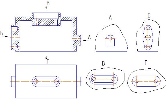
The rules for depicting objects (products, structures and their component elements) on drawings for all industries and construction are established by GOST 2.305 - 2008* “Images - views, sections, sections.”
Images of objects must be made using the rectangular (orthogonal) projection method. In this case, the object is placed between the observer and the corresponding projection plane. When constructing images of objects, the standard allows the use of conventions and simplifications, as a result of which the specified correspondence is violated. Therefore, the resulting figures when projecting an object are called not projections, but images. The faces of a hollow cube are taken as the main projection planes, into which an object is mentally placed and projected onto the inner surfaces of the faces. The faces are aligned with the plane (Figure 2.1). As a result of this projection, the following images are obtained: front view, top view, left view, right view, rear view, bottom view.
The image on the frontal plane is taken as the main one in the drawing. The object is positioned relative to the frontal plane of projections so that the image on it gives the most complete idea of the design features of the object and its functional purpose.
Let's consider main image selection using the example of an object such as a chair. Let us depict its projections schematically:
Let's think: the functional purpose of the object is to sit on it. In which of the figures this purpose is most clear - probably this is figure 1 or 2, the 3rd is the least informative.
The design features of the item include a seat itself, a backrest for the convenience of sitting on a chair, located at a certain angle relative to the seat, legs that position the seat at a certain distance from the floor. Which of the figures shows these features most clearly? Obviously this is Figure 1.
Conclusion - we choose projection number 1 as the main view, as it is the most informative and provides the most complete information about the functional purpose of the chair and its design features.
It is necessary to think in a similar way when choosing the main image of any subject!
The images in the drawing, depending on their content, are divided into types, sections, sections.
View - image of the visible part of the surface of an object facing the observer.
Types are divided into basic, local and additional.
Main types — images are obtained by projecting an object onto a projection plane. There are six of them in total, but more often than others, I use the main three to obtain information about the subject: horizontal π 1, frontal π 2 and profile π 3 (Figure 2.1). With this projection we get: front view, top view, left view.
The names of views on the drawings are not inscribed if they are located in a projection relationship (Figure 2.1). If the views from above, to the left and to the right are not in projection connection with the main image, then they are marked on the drawing with an inscription of type “A”. The direction of view is indicated by an arrow, indicated by a capital letter of the Russian alphabet. When there is no image that can show the direction of view, the name of the species is inscribed.


Figure 2.1 Formation of main species
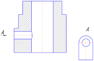
Local view - an image of a separate limited area of the surface of an object on one of the main projection planes. The local view can be placed in any free space of the drawing, marked with an inscription like “A”, and the associated image of the object should have an arrow indicating the direction of view, with the corresponding letter designation (Figure 2.2 a, b).
 |
| A |
 |
| b |
Figure 2.2 – Local species
The local species may be limited to the cliff line, in the smallest possible size (Figure 2.2, a), or not limited (Figure 2.2, b).
Additional views— images obtained on planes non-parallel to the main planes of projections. Additional views are performed in cases where any part of the object cannot be shown in the main views without distorting its shape and size. The additional view is marked on the drawing with an inscription of type “A” (Figure 2.3, a), and an arrow with the corresponding letter designation is placed next to the additional view of the image of the object (Figure 2.3, a), indicating the direction of view.
When an additional view is located in direct projection connection with the corresponding image, the arrow and inscription above the view are not applied (Figure 2.3, b). The secondary view can be rotated while maintaining the same position as the item in the main image. In this case, a sign (“Rotated”) is added to the inscription “A” (Figure 2.3, c).
Basic, local and additional views are used to depict the shape of the external surfaces of an object. A successful combination of them allows you to avoid dashed lines, or reduce their number to a minimum. To reduce the number of images, it is allowed to show the necessary invisible parts of the surface in views using dashed lines. However, identifying the shape of the internal surfaces of an object using dashed lines significantly complicates reading the drawing, creates preconditions for its incorrect interpretation, and complicates the application of dimensions and symbols, so their use should be limited and justified. To identify the internal (invisible) configuration of an object, conventional images are used - cuts and sections.
Figure 2.3
2.2 Sections
A section is an image of an object mentally dissected by one or more planes.
The section shows what is located in the secant plane and what is located behind it.
2.2.1 Classification of cuts
Depending on the number of cutting planes The sections are divided into (Figure 2.4):
- simple— with one cutting plane (Figure 2.6);
- complex— with several cutting planes (Figure 2.9, 2.10).

Figure 2.4 - Classification of cuts
The position of the cutting plane is shown in the main image with a thick open line (1.5s, where s– thickness of the main line). The length of each stroke is from 8 to 20 mm. The direction of view is shown by arrows perpendicular to the strokes. Arrows are drawn at a distance of 2-3 mm from the outer ends of the strokes. The name of the cutting plane is indicated in capital letters of the Russian alphabet. The letters are applied parallel to the horizontal lines of the main inscription, regardless of the position of the arrows (Figures 2.5, 2.6, 2.9, 2.10, 2.11).
If, when making a simple cut that is in projection connection with the main image, the cutting plane coincides with the plane of symmetry, then the cutting plane is not depicted and the cut is not labeled.

Figure 2.5 – Designations of sections in the drawing

Figure 2.6 - Simple section: a) - frontal; b) - local
Depending on the cutting plane position relative to the horizontal plane of projections, the sections are divided into:
- horizontal — the secant plane is parallel to the horizontal plane of projections (Figure 2.7, b);
- vertical – the secant plane is perpendicular to the horizontal plane of projections (Figure 2.7, c, d);
- inclined– the secant plane makes an angle with the horizontal projection plane that is different from the right angle (Figure 2.8).

Figure 2.7 a – Model of the “Crank” part


Figure 2.7 b - Simple horizontal section
Vertical the cuts are called:
- frontal , if the cutting plane is parallel to the frontal plane of projections (Figure 2.7, c);
- profile, if the cutting plane is parallel to the profile plane of projections (Figure 2.7, d).


Figure 2.7 c – Simple frontal section


Figure 2.7 d - Simple profile section

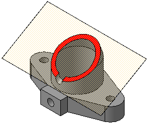
Figure 2.8 – Oblique section
Complex cuts are divided into:
- stepped , if the cutting planes are parallel (stepped horizontal, stepped frontal) (Figure 2.9);
- broken lines, if the cutting planes intersect (Figure 2.10).

Figure 2.9 - Complex - Stepped cut

Figure 2.10 - Complex - Broken cut
The cuts are called:
- longitudinal, if the cutting planes are directed along the length or height of the object (Figure 2.7, c);
- transverse, if the cutting planes are directed perpendicular to the length or height of the object (Figure 2.7, d).
Sections that serve to clarify the structure of an object only in certain, limited places are called local .


Figure 2.11 a - Examples of making cuts

Figure 2.11 b - Examples of making sections combined with views
2.2.2 Making cuts
Horizontal, frontal and profile sections can be located in place of the corresponding main views (Figure 2.11, a, b).
Part of the view and part of the corresponding section can be connected by separating them with a solid wavy line or a line with a break (Figure 2.11, b). It should not coincide with any other lines in the image.
If half of the view and half of the section are connected, each of which is a symmetrical figure, then the dividing line is the axis of symmetry (Figures 2.11, b; 2.12). You cannot connect half a view with half a section if any line of the image coincides with the axial line (for example, an edge). In this case, connect a larger part of the view with a smaller part of the section, or a larger part of the section with a smaller part of the view.
It is allowed to separate the section and the view by a thin dash-dotted line coinciding with the trace of the plane of symmetry not of the entire object, but only of its part, if it represents a body of rotation. When connecting half of the view with half of the corresponding section, the section is located to the right of the vertical axis and below the horizontal (Figure 2.12).

Figure 2.12

Figure 2.13
Local cuts are highlighted in the view as solid wavy lines. These lines should not coincide with any other lines in the image (Figure 2.13).
Sectional figures obtained by different cutting planes when performing complex cut, do not separate one from the other by any lines.
A complex stepped section is placed in place of the corresponding main view (Figure 2.9) or anywhere in the drawing.
With broken cuts, the secant planes are conventionally rotated until they align into one plane, and the direction of rotation may not coincide with the direction of view. If the combined planes turn out to be parallel to one of the main projection planes, then the broken section can be placed in the place of the corresponding type (Figure 2.10).
When rotating the cutting plane, the elements of the object located behind it are drawn as they are projected onto the corresponding plane with which the alignment is made. It is allowed to connect a stepped cut with a broken one in the form of one complex cut.
2.3 Sections
Section called the image of a figure obtained by mentally dissecting an object with a cutting plane(Figure 2.14).
The section shows only what falls directly into the cutting plane.
The cutting planes are chosen so as to obtain normal cross sections.
Sections are divided into:
- sections included in the section (Figure 2.15, a);
- sections not included in the section Figure 2.15.b).
Sections not included in the composition are divided into:
- issued(Figures 2.14, a; 2.14, c; 2.15, b; 2.16, a; 2.17, a; 2.18);
- superimposed(Figures 2.14, b; 2.16, b; 2.17, b).
Extended sections are preferable and they can be located in the gap between parts of the same type, on the continuation of the trace of the cutting plane with a symmetrical section figure, at any place in the drawing field, as well as with a rotation (Figures 2.14, a, c; 2.15, b; 2.16, a; 2.17, a);
To depict the trace of the cutting plane in the drawing, use a thick open line with arrows indicating the direction of view, and designate the cutting plane in capital letters of the Russian alphabet. The section is accompanied by an inscription of type AA (Figure 2.14).
The ratio of the sizes of the arrows and the strokes of the open line must correspond to Figure 2.14. The starting and ending strokes must not intersect the outline of the image.
Letter designations are assigned in alphabetical order without repetition and, as a rule, without gaps. The font size of the letter designations should be approximately two times larger than the size of the digits of the size numbers. The letter designation is located parallel to the main inscription, regardless of the position of the cutting plane.
In the general case, when the section is located in any free space in the drawing, the position of the trace of the cutting plane is depicted as indicated above, and the image of the section is accompanied by an inscription corresponding to the name of the cutting plane (Figure 2.14, a; 2.15, b).
In the cases shown in Figures: 2.14, b, c; 2.17, a, b; 2.18, a (superimposed sections; sections made in a break in the view; sections made on the continuation of the trace of the cutting plane) - for symmetrical sections the trace of the cutting plane is not depicted and the section is not accompanied by an inscription.

Figure 2.14 A

Figure 2.14 b

Figure 2.14 V
For asymmetrical sections , located in a gap, or superimposed, the trace of the cutting plane is depicted, but not accompanied by letters (Figure 2.16). The section is also not accompanied by an inscription.
The outline of the extended section is drawn with a thick solid line (the main line), and the outline of the superimposed section is drawn with a thin solid line, while the outline of the view is not interrupted.
 |
|
| A | b |
Figure 2.15
 |
|
| A | b |
Figure 2.16

Figure 2.17 A,b
 |
|
| A | b |
Figure 2.18
For several identical sections of the same object, the section lines are designated by one letter and one section is drawn. If the cutting planes are directed at different angles, then the “Rotated” sign is not applied (Figure 2.19).

In a longitudinal section, the following root zones (plant root areas) are distinguished:
- Growth zone with root cap;
- zone of elongation and beginning of cell differentiation;
- suction zone;
- conductive zone.
Root zones
Growth
The growth zone (division zone) of the root occupies the tip 2-3 mm long. This is a zone of actively dividing cells, the root meristem. All root tissues arise from this educational tissue.
Growth area covered root cap, which protects it from damage and facilitates the advancement of the root in the soil. The cells of the cap have increased turgor. As the roots deepen in the soil, they are erased, their outer layer is peeled off, and new cells grow from the inside due to the root meristem.
Sprains
In the elongation zone, the cells greatly increase in the longitudinal direction and become cylindrical. Large vacuoles appear in them. The combined growth of cells in this zone creates a force that forces the root deeper into the soil.
This zone is also small and occupies a few millimeters. In its upper part, cells begin to specialize, finally turning into vessels, tracheids and other types of root cells in the suction zone.
Suction
The root absorption zone ranges in length from a few millimeters to several centimeters. Its surface is protected by integumentary tissue - skin with root hairs. Under the skin is the root bark, surrounding its central part with a conducting system.
The conductive zone is the entire rest of the root, from the suction zone to the plant stem. This area has a denser integumentary tissue, is thickened, the number of vessels and sieve tubes is increased due to the activity of the cambium.
The root conduction zone is an intermediary between the suction zone and the above-ground part of the plant.
Summary table of the structure and functions of root zones
| Zone name | Structural features | Functions |
|---|---|---|
| Division zone | Small living cells that divide quickly | The beginning of all other zones and root tissues |
| Growth zone | Cells grow and increase in size | Provides basic root growth |
| Suction zone | The outer layer is represented by cells with root hairs | Provides absorption of water with beneficial substances dissolved in it |
| Venue area | Conductive tissues are well developed | Transport |
Internal structure of a plant root
Outer covering tissue of the root - skin- differs from the skin of the stem and leaf in the presence of root hairs, the absence of stomata and cuticle, easy permeability to water, and absorption capacity.
The skin cells are arranged in one layer. Many of them have root hairs - elongated cylindrical outgrowths of the outer wall of skin cells, ranging from 0.15 mm to 1 cm in length with a diameter of hundredths of a millimeter. The cell nucleus passes into the root hair and is usually located at its very end.
In addition to the nucleus, the cytoplasm of the root hair contains vacuoles with cell sap and colorless plastids. The surface of the hairs is covered with a mucous substance that glues them to soil particles.
Root hairs short-lived. They are formed in 30-40 hours, live for 10-20 days, then die off. To replace them, new ones are formed in the young part of the root, and the area with dead hairs becomes a conductive zone. The number of root hairs per 1 mm2 reaches several hundred (for example, in corn - 425, in peas - 230). Thanks to their presence, the suction surface of the root increases tens of times.
Root bark, adjacent to the skin from the inside, consists of cells of the main tissue arranged in several rows. Cortical cells have different sizes. Directly under the skin they are large, and in deeper layers they are smaller.
The innermost layer of the bark ( endoderm), enclosing the central part of the root (central cylinder) with a conducting system, consists of one row of densely packed cells. Their outer walls (from the bark side) are thin, while the lateral and internal walls are thickened and impermeable to water and gases.
Between the thick-walled cells there is a small number of thin-walled cells located opposite the vessels of the central cylinder. These are passage cells; they conduct water from the root bark into the vessels of the central cylinder.
Central cylinder occupies the middle part of the stem and consists of various tissues. Its outer layer, adjacent to the endoderm from the inside, consists of thin-walled parenchyma cells and is called the pericycle, or root layer.
Pericycle cells (secondary educational tissue) periodically divide and give rise to lateral roots, root parenchyma, adventitious buds of root shoots, and cambium.
Further, towards the center of the axial cylinder, there is a closed vascular-fibrous bundle, in which sections of phloem and xylem alternate, located radially. The center of the axial cylinder of the root in most plant species is occupied by one large or several small vessels. In some species, the center is occupied by cells of the main tissue (parenchyma), which also fills the gaps between the phloem and xylem areas.
Root Anatomy (Part 2)
Primary root structure can be viewed under a microscope on a cross-section of the suction zone of a young root. A similar preparation shows that the root consists of the epidermis (epiblema), which forms root hairs, primary root cortex, located under the epidermis, occupying the main part of the root and consisting of cells of the main tissue. The inner part of the root is called central cylinder, which consists mainly of conductive tissues (Fig. 2).
Fig.2. Cross sections of the root:
I - the incision is made in the area of root hairs, the epidermis with numerous root hairs, the main cortex tissue and the central cylinder are visible. II - central root cylinder: a - a large vessel, from which five rays of smaller vessels diverge, with areas of phloem (phloem) between them; b - endodermal cells; c - passage cells, d - pericycle, or root layer.The main tissue of the root cortex cells contains protoplast, as well as reserve substances, crystals, resins, etc. The innermost layer of the cortex forms endodermis, which surrounds the central cylinder and consists of several elongated cells. On cross sections, the radial membranes of these cells have dark spots or very thickened internal and lateral lignified membranes that do not allow water to pass through. Among them there are vertical rows access cells with thin-walled cellulose membranes, they are located opposite the wood vessels and serve to pass water and salts flowing from the root hairs through the bark cells into the wood vessels.
Located inside the endodermis central cylinder, the outer layer of which is called root layer(pericycle), since lateral roots develop from it, which then grow through the bark and come out. Lateral roots are usually formed opposite the rays of the wood, and therefore they are distributed on the root in regular rows according to the number of rays of the wood or twice as many rows.
The central cylinder contains conductive tissue, consisting of aquifers - tracheas and tracheids, forming wood (xylem), and sieve tubes with accompanying cells, forming phloem (phloem) and conducting organic matter. Since the primary wood at the root is located in the form of rays, the number of which varies (from 2 to 20), then areas of primary phloem are distributed in the spaces between the rays of the primary wood and their number corresponds to the number of rays of the wood.
Tracheas, or vessels, are hollow tubes whose walls have various thickenings. Tracheids are elongated (prosenchymal) dead cells with pointed ends.
Through the tracheae and tracheids, water and dissolved salts rise up the root and further along the stem, and through the sieve tubes of the bast, organic substances (sugar, protein substances, etc.) descend from the stem down into the root and into its branches.
The mechanical elements of bast and wood (bast fibers and wood fibers) are distributed between the cells of the conductive tissue. Living parenchyma cells are also found in the central cylinder of the root.
In the roots monocots changes during life are reduced only to the death of root hairs and the suberization of cells of the outer cortex, to the appearance of mechanical tissues. Only in tree-like monocots with thickening roots and trunks (dracaenas, palms) does a cambium appear and secondary changes occur.
U dicotyledonous plants already during the first year of life, the primary structure of the root described above undergoes sharp secondary changes associated with the fact that a strip of cambium appears between the primary wood (xylem) and the primary phloem; if its cells are deposited inside the root, they turn into secondary wood (xylem), and outward into secondary bast (phloem). Cambium cells arise from parenchyma cells located between the primary wood and the phloem. They are divided by tangential partitions (Fig. 3).
Fig.3. The beginning of secondary changes in the root of a dicotyledonous plant (common bean):
1 - main cortex tissue; 2 - endoderm; 3 - root layer (pericycle); 4 - cambium; 5 - bast (phloem); 6 - primary xylem.Pericycle cells, located opposite the rays of wood, divide, forming parenchymal tissue, which turns into core beam. The remaining cells of the pericycle, which are the outer layer of the central cylinder of the root, also begin to divide along their entire length, and from them cork tissue arises, separating the inner part of the root from the primary cortex, which gradually dies and is shed from the root.
Cambial layer closes around the primary wood of the central cylinder, and as a result of the division of its cells, secondary wood grows inside, and a continuous bast is formed towards the periphery, moving further and further from the primary wood. The cambium initially looks like a curved line, and later flattens out and takes the shape of a circle.
In autumn and winter, cambium cell division ceases, and in spring it begins with renewed vigor. As a result, layers of wood are formed in perennial roots, and the root becomes similar in structure to the stem. You can distinguish roots from stems by the primary wood remaining in the center of the root in the form of radial rays(Fig. 2). In the root, the pith rays rest against the primary wood, whereas in the stem they always rest against the pith.
The vessels of the wood and the sieve tubes of the bast pass from the root directly into the stem, where they are located not in radial rays, as in the primary structure of the root, but in the form of ordinary closed (monocots) and open (dicots) vascular-fibrous bundles. Regrouping of wood and bast occurs in the root collar in the subcotyledon.
The structure of the root of a plant is studied by the science of botany. Studying this material will help you learn the characteristics of this part of the plant.
What is a root
The root is a constantly growing and developing organ. Its most important function is to carry out the growth and vital activity of the plant. This includes nutrition and respiratory function. Its length and shape constantly changes as the stem grows.
Inside this organ are all the vitamins and substances that are obtained and formed through synthesis.
Root zones
Detailed tables describing the zones of the root system can be found in textbooks on botany. We will tell you the main points.
In the structure of the root system, important zones are distinguished from the top to the tail. The root cap serves as a cover for the tail part and protects the end from damage. With each growth of the end of the root, one can observe the resulting wrinkling of the cap and the appearance of new cells.
Below the cover there is a division zone. This is where cell reproduction occurs. The length of this zone is usually only a few millimeters. Above it is a growth zone in which these cells elongate.
Next comes the suction zone. Its length is about one centimeter. This is where seedlings form. They are called root hairs. All of them are clearly visible to the naked eye, and together they form a thin white fluff on the spine. Root hairs consist of a nucleus, a membrane, leukocytes and cytoplasm.
The suction zone provides fluid and mineral nutrition. Root hairs penetrate between soil cells and absorb nutrition. Next, the nutrients move through the internal cells of the root to the conduction zone. This zone carries out the transition of the necessary important nutrients to the cells of the stem.
There is a continuous relationship between the root and the stem. From the stem, all the organic nutrients necessary for its growth enter the root. The conduction system zone is also located at the root tip. With the help of fibers, interaction occurs between the elements of the root.
Root modifications
To survive in different conditions, plants can have completely different types of roots. The characteristics of the ivy plant help it climb any height with the help of roots-trailers.
Roots found in rutabaga, turnips, and carrots. These are mainly biennial plants. If a person needs to get seeds, then the fruit is left for next year. But mostly they eat root vegetables.
Root tubers found in lilies, dahlias and other flowers. They accumulate all the nutrients needed for nutrition. They are formed from lateral or adventitious roots.
Support roots found in many tropical trees. They protrude from the soil, creating columnar supports for plants. For example, the banyan plant, some types of ficus.
Aerial roots have orchids and other tropical flowers. The growth and life of a plant occurs when the hanging roots draw in water and nutrition from the air sphere.
Sucker roots found in many poisonous plants. With their help, they attach themselves to other plants, sucking nutrients and moisture from them.
Types of roots
In biology there are three types of roots:
- Subordinate clauses are called shoots directed horizontally, parallel to the soil. They originate from various organs of the plant: on stems, leaves, and the main root.
- main root usually the largest, goes down into the ground, grows vertically downwards. It grows from an embryonic seed.
- Lateral can grow on both adventitious roots and the main root.
Types of root systems
There are two types of root systems: fibrous And core. The taproot type structure consists of a basic tap root. He is strong and well developed.
The fibrous type consists of several identical processes that intertwine with each other and are shaped like a nest or a bundle.
Internal structure of the root
Let's examine the microscopic structure of the root system in a cross section using a drawing with captions. A longitudinal section can show how the root is structured inside.
The root has several layers:
- peel;
- primary cortex;
- tissue forming the outer layer;
- conductive fabrics;
- vessels through which nutrients, minerals and water move;
- tissue that stores nutrients.
Conclusion
We figured out what roots come in shape and type, what they serve for plants, and what important role they play. By studying the anatomical structure of the root system, you can find out its meaning and function.


